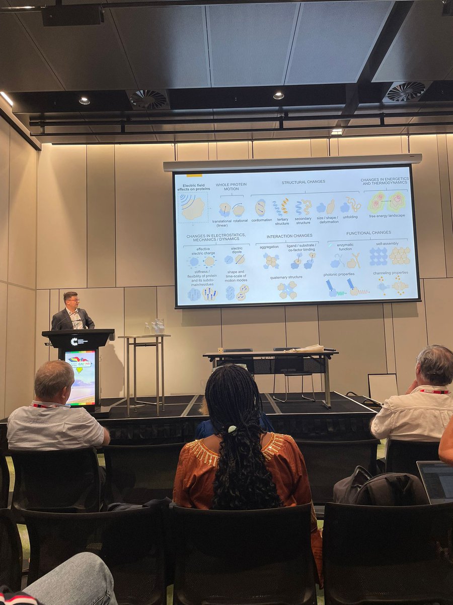
Bioelektrodynamika: O týmu

🔊🔬⚡We present a fast and accurate method to extract complex permittivity from broadband S-parameters—validated up to 50 GHz and ideal for real-time, noninvasive analysis of tiny protein samples. Big kudos to Petr Kurka and all the authors for their exceptional work.🥇👏
🔎👇

🔊🔬⚡We present a fast and accurate method to extract complex permittivity from broadband S-parameters—validated up to 50 GHz and ideal for real-time, noninvasive analysis of tiny protein samples. Big kudos to Petr Kurka and all the authors for their exceptional work.🥇👏
🔎👇
Bioelectrodynamics @BioED_IPE

🔊🔬⚡In our recently published work, we present a fast, broadband method to extract complex #dielectric #permittivity from μL-scale #biomolecular samples using a CBCPW. It offers accurate, sample-efficient characterization for #biomedical #research. 🔎📖👉https://ieeexplore.ieee.org/document/10973163

🎨A little glow, a little competition, and a team that always brings the fun 🥇⛳️🤩
#TeamOuting #NeonMinigolf #WorkCrew #BrightMoments #TeamEnergy

🔊🔬⚡we discovered that calibrating our permittivity-extraction method at ~75% of the biomolecular concentration range gives the best accuracy. It captures the linear S-parameter behavior while avoiding nonlinear distortion—improving consistency across samples. 🔎📖👇

🔊🔬⚡we discovered that calibrating our permittivity-extraction method at ~75% of the biomolecular concentration range gives the best accuracy. It captures the linear S-parameter behavior while avoiding nonlinear distortion—improving consistency across samples. 🔎📖👇
Bioelectrodynamics @BioED_IPE

🔊🔬⚡In our recently published work, we present a fast, broadband method to extract complex #dielectric #permittivity from μL-scale #biomolecular samples using a CBCPW. It offers accurate, sample-efficient characterization for #biomedical #research. 🔎📖👉https://ieeexplore.ieee.org/document/10973163

Great news from our recent Czech Academy of Sciences evaluation: our BioED team earned exceptional marks. Most of our outputs were rated outstanding, and our PEF lab-on-chip, microwave microfluidic sensing, and BAL imaging platforms were highlighted as unique, high-impact tech.👏

Load More

Výpočetní modelování: elektromagnetické vlastnosti biologických systémů

Jak biologické systémy reagují na a generují elektromagnetické pole? Používáme molekulární i hrubozrnné modelování a simulace k směrování a interpretaci našich experimentů odpovídající na tuto otázku. V současné době se zaměřujeme na modelování účinků intenzivních elektrických pulzů na struktury cytoskeletu. Dále používáme klasickou molekulární dynamiku pro simulaci dielektrických vlastností biologické hmoty. Naše práce zahrnuje i modelování procesů, které vytvářejí endogenní biologické elektromagnetické pole v mikrovlnném a viditelném pásmu.

Publikace:

  1. Průša and Cifra, “Electro-detachment of kinesin motor domain from microtubule in silico,” Comput Struct Biotechnol J, vol. 21, 1349–1361, 2023.
  2. Vahalová and Cifra, “Biological autoluminescence as a perturbance-free method for monitoring oxidation in biosystems,” Progress in Biophysics and Molecular Biology, vol. 177, pp. 80–108, 2023.
  3. Průša et al. “Electro-Opening of a Microtubule Lattice in Silico.” Computational and Structural Biotechnology Journal 19 (2021): 1488–96.
  4. Marracino et al. “Tubulin Response to Intense Nanosecond-Scale Electric Field in Molecular Dynamics Simulation.” Scientific Reports 9, no. 1 (2019): 10477.
  5. Průša and Cifra. “Dependence of Amino-Acid Dielectric Relaxation on Solute-Water Interaction: Molecular Dynamics Study.” Journal of Molecular Liquids 303 (2020): 112613.
  6. Cifra and Pospišil. “Ultra-Weak Photon Emission from Biological Samples: Definition, Mechanisms, Properties, Detection and Applications.” Journal of Photochemistry and Photobiology B: Biology 139 (2014): 2–10.
  7. Cifra et al. “Biophotons, Coherence and Photocount Statistics: A Critical Review.” Journal of Luminescence 164 (2015): 38–51.
  8. Kucera et al. “Spectral Perspective on the Electromagnetic Activity of Cells.” Current Topics in Medicinal Chemistry 15, no. 6 (2015): 513–522.
  9. Havelka et al. “Multi-Mode Electro-Mechanical Vibrations of a Microtubule: In Silico Demonstration of Electric Pulse Moving along a Microtubule.” Applied Physics Letters 104, no. 24 (2014): 243702.
  10. Havelka et al. “Electro-Acoustic Behavior of the Mitotic Spindle: A Semi-Classical Coarse-Grained Model.” PLoS ONE 9, no. 1 (2014): e86501.
  11. Havelka et al “High-Frequency Electric Field and Radiation Characteristics of Cellular Microtubule Network.” Journal of Theoretical Biology 286 (2011): 31–40.
  12. Cifra et al. “Electric Field Generated by Axial Longitudinal Vibration Modes of Microtubule.” Biosystems 100, no. 2 (2010): 122–31.

Elektromagnetické čipy: návrh a technologie

Jaké nástroje používáme k ověření našich teorií a modelů? Vyvíjíme jedinečné radiofrekvenční/mikrovlnné čipy a planární struktury včetně mikrofluidiky pro ověření skrze experimenty. Čipy jsou navrženy pomocí moderních výpočetních nástrojů a jsou vyráběny pomocí technik mikro- a nanofabrikace. Naše čipy jsou navrženy tak, aby byly kompatibilní s pokročilými mikroskopickými systémy pro pozorování biomolekulárních struktur a buněk ve chvíli, kdy jsou vystaveny elektromagnetickému poli.

Biofyzikální experimenty: elektromagnetické vlastnosti proteinů a buněk

Teoretické modely a simulace ověřujeme skrze experimenty: v současné době se zaměřujeme na experimenty na nanostrukturách cytoskeletálních proteinů, konkrétně na mikrotubulech a s nimi souvisejících proteinech. Mikrotubuly jsou klíčové struktury nacházející se v každé buňce a umožňující dělení buněk a intracelulární transport. Naše současné projekty se zaměřují na měření elektromagnetických/dielektrických vlastností mikrotubulárních systémů a jejich řízení pomocí ultrakrátkých elektrických a elektromagnetických pulzů. Naše experimentální práce také zahrnuje měření a analýzu endogenního světla (luminiscence) buněk, v současné době pro monitorování oxidativních biologických vlivů elektromagnetického pole.

Publikace:

  1. Vahalová et al. “Biochemiluminescence Sensing of Protein Oxidation by Reactive Oxygen Species Generated by Pulsed Electric Field,” Sens Actuators B Chem, vol. 385, p. 133676, 2023.
  2. Poplová et al. “Biological Auto(chemi)luminescence Imaging of Oxidative Processes in Human Skin,” Analytical Chemistry, vol. 95, no. 40, pp. 14853–14860, 2023.
  3. Havelka et al. “Nanosecond Pulsed Electric Field Lab-on-Chip Integrated in Super-Resolution Microscope for Cytoskeleton Imaging.” Advanced Materials Technologies 5, no. 3 (2020): 1900669.
  4. Chafai et al. “Reversible and Irreversible Modulation of Tubulin Self‐Assembly by Intense Nanosecond Pulsed Electric Fields.” Advanced Materials 31, no. 39 (2019): 1903636.
  5. Krivosudský et al “Resolving Controversy of Unusually High Refractive Index of a Tubulin.” EPL (Europhysics Letters) 117, no. 3 (2017): 38003.
  6. Burgos et al. “Ultra-Weak Photon Emission as a Dynamic Tool for Monitoring Oxidative Stress Metabolism.” Scientific Reports 7, no. 1 (2017): 1229.
  7. Poplová et al. “Poisson Pre-Processing of Nonstationary Photonic Signals: Signals with Equality between Mean and Variance.” PLOS ONE 12, no. 12 (2017): e0188622.
  8. Saeidfirozeh et al. “Endogenous Chemiluminescence from Germinating Arabidopsis Thaliana Seeds.” Scientific Reports 8, no. 1 (2018): 16231.
  9. Sardarabadi et al. “Enhancement of the Biological Autoluminescence by Mito-Liposomal Gold Nanoparticle Nanocarriers.” Journal of Photochemistry and Photobiology B: Biology 204 (2020): 111812.
Skip to content