Bioelektrodynamika: O týmu

📣📰#Protein dipoles can act as tiny handles for electric-field-driven molecular orientation. Our review explores this underutilized effect—including “orientation before destruction”—and its exciting potential for single-particle X-ray imaging. 🔎👉

📣📰#Protein dipoles can act as tiny handles for electric-field-driven molecular orientation. Our review explores this underutilized effect—including “orientation before destruction”—and its exciting potential for single-particle X-ray imaging. 🔎👉
Bioelectrodynamics @BioED_IPE

🚨⚡📰In our recently published review, we explore how #ElectricFields can modulate #protein structure, dynamics, and function—offering a reagent-free physical approach with exciting potential across #biomedicine and #biotechnology. 🔎👇
https://shorturl.at/dfsBm
@NeuronColl


🚨📰Can we control the molecular machines of life with electric fields? In our recent review, we highlight how EFs can influence protein motion, structure, interactions and function—and explore their potential as a reagent-free tool for protein control.🔎👉

🚨📰Can we control the molecular machines of life with electric fields? In our recent review, we highlight how EFs can influence protein motion, structure, interactions and function—and explore their potential as a reagent-free tool for protein control.🔎👉
Bioelectrodynamics @BioED_IPE

🚨⚡📰In our recently published review, we explore how #ElectricFields can modulate #protein structure, dynamics, and function—offering a reagent-free physical approach with exciting potential across #biomedicine and #biotechnology. 🔎👇
https://shorturl.at/dfsBm
@NeuronColl

🚨⚡📰In our recently published review, we explore how #ElectricFields can modulate #protein structure, dynamics, and function—offering a reagent-free physical approach with exciting potential across #biomedicine and #biotechnology. 🔎👇
https://shorturl.at/dfsBm
@NeuronColl
📢 Conference Alert!🚨
@mcer33 and Přemysl Klajs are set to attend #URSIGASS2026 in Kraków! 🇵🇱
They’ll be representing our group, sharing our research, and connecting with the international #RadioScience community.
Wishing them an inspiring and successful #conference! 🧑‍🏫🖖

Load More

Výpočetní modelování: elektromagnetické vlastnosti biologických systémů

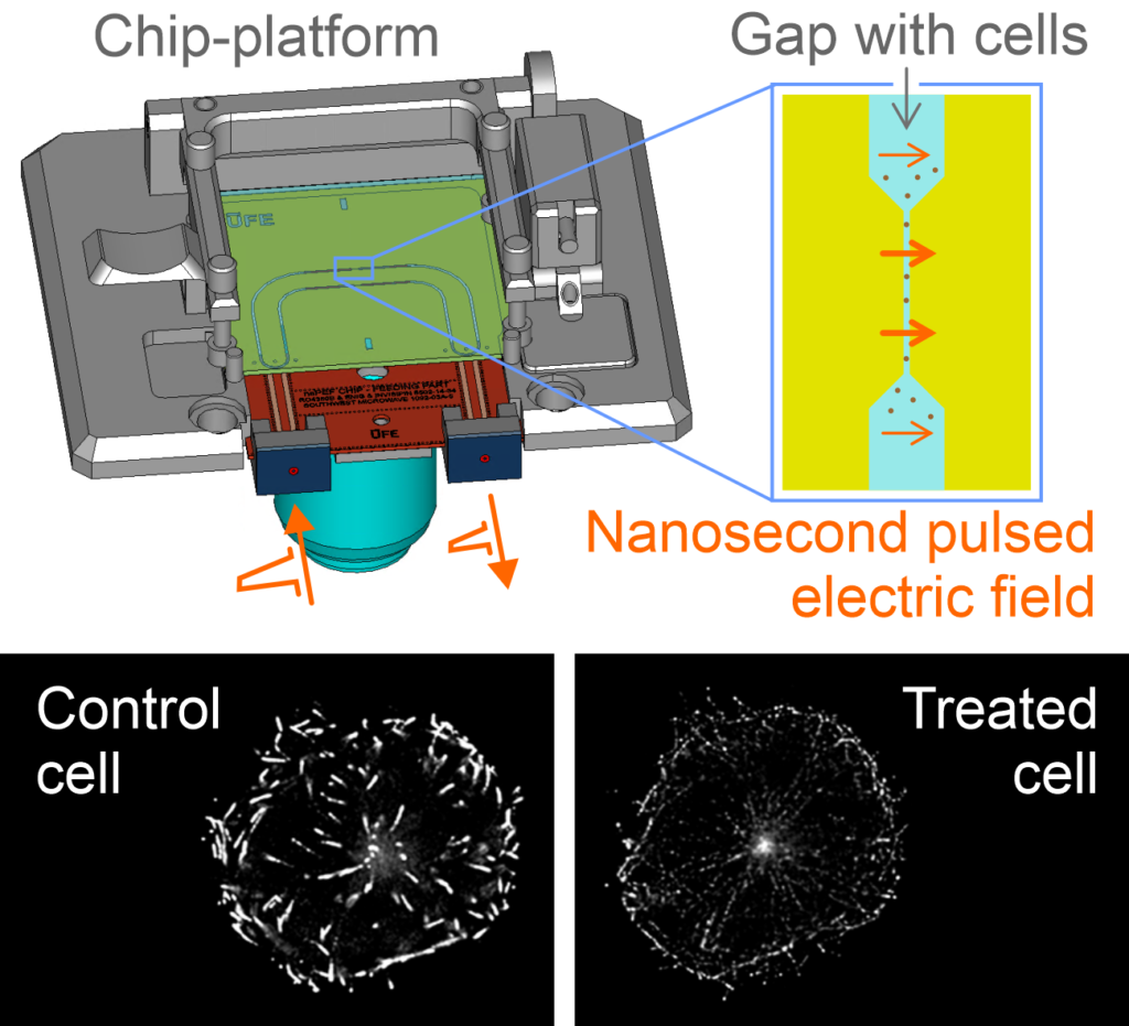
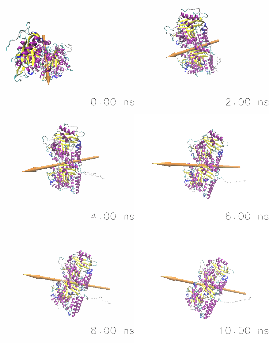
Jak biologické systémy reagují na a generují elektromagnetické pole? Používáme molekulární i hrubozrnné modelování a simulace k směrování a interpretaci našich experimentů odpovídající na tuto otázku. V současné době se zaměřujeme na modelování účinků intenzivních elektrických pulzů na struktury cytoskeletu. Dále používáme klasickou molekulární dynamiku pro simulaci dielektrických vlastností biologické hmoty. Naše práce zahrnuje i modelování procesů, které vytvářejí endogenní biologické elektromagnetické pole v mikrovlnném a viditelném pásmu.

Publikace:

  1. Průša and Cifra, “Electro-detachment of kinesin motor domain from microtubule in silico,” Comput Struct Biotechnol J, vol. 21, 1349–1361, 2023.
  2. Vahalová and Cifra, “Biological autoluminescence as a perturbance-free method for monitoring oxidation in biosystems,” Progress in Biophysics and Molecular Biology, vol. 177, pp. 80–108, 2023.
  3. Průša et al. “Electro-Opening of a Microtubule Lattice in Silico.” Computational and Structural Biotechnology Journal 19 (2021): 1488–96.
  4. Marracino et al. “Tubulin Response to Intense Nanosecond-Scale Electric Field in Molecular Dynamics Simulation.” Scientific Reports 9, no. 1 (2019): 10477.
  5. Průša and Cifra. “Dependence of Amino-Acid Dielectric Relaxation on Solute-Water Interaction: Molecular Dynamics Study.” Journal of Molecular Liquids 303 (2020): 112613.
  6. Cifra and Pospišil. “Ultra-Weak Photon Emission from Biological Samples: Definition, Mechanisms, Properties, Detection and Applications.” Journal of Photochemistry and Photobiology B: Biology 139 (2014): 2–10.
  7. Cifra et al. “Biophotons, Coherence and Photocount Statistics: A Critical Review.” Journal of Luminescence 164 (2015): 38–51.
  8. Kucera et al. “Spectral Perspective on the Electromagnetic Activity of Cells.” Current Topics in Medicinal Chemistry 15, no. 6 (2015): 513–522.
  9. Havelka et al. “Multi-Mode Electro-Mechanical Vibrations of a Microtubule: In Silico Demonstration of Electric Pulse Moving along a Microtubule.” Applied Physics Letters 104, no. 24 (2014): 243702.
  10. Havelka et al. “Electro-Acoustic Behavior of the Mitotic Spindle: A Semi-Classical Coarse-Grained Model.” PLoS ONE 9, no. 1 (2014): e86501.
  11. Havelka et al “High-Frequency Electric Field and Radiation Characteristics of Cellular Microtubule Network.” Journal of Theoretical Biology 286 (2011): 31–40.
  12. Cifra et al. “Electric Field Generated by Axial Longitudinal Vibration Modes of Microtubule.” Biosystems 100, no. 2 (2010): 122–31.

Elektromagnetické čipy: návrh a technologie

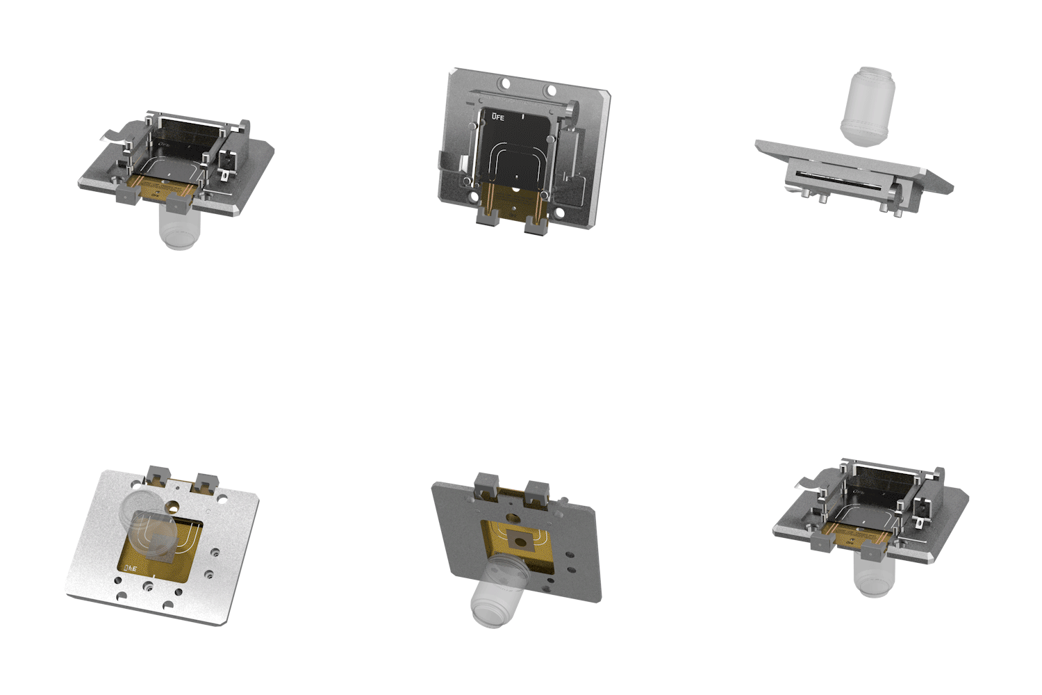
Jaké nástroje používáme k ověření našich teorií a modelů? Vyvíjíme jedinečné radiofrekvenční/mikrovlnné čipy a planární struktury včetně mikrofluidiky pro ověření skrze experimenty. Čipy jsou navrženy pomocí moderních výpočetních nástrojů a jsou vyráběny pomocí technik mikro- a nanofabrikace. Naše čipy jsou navrženy tak, aby byly kompatibilní s pokročilými mikroskopickými systémy pro pozorování biomolekulárních struktur a buněk ve chvíli, kdy jsou vystaveny elektromagnetickému poli.

Biofyzikální experimenty: elektromagnetické vlastnosti proteinů a buněk

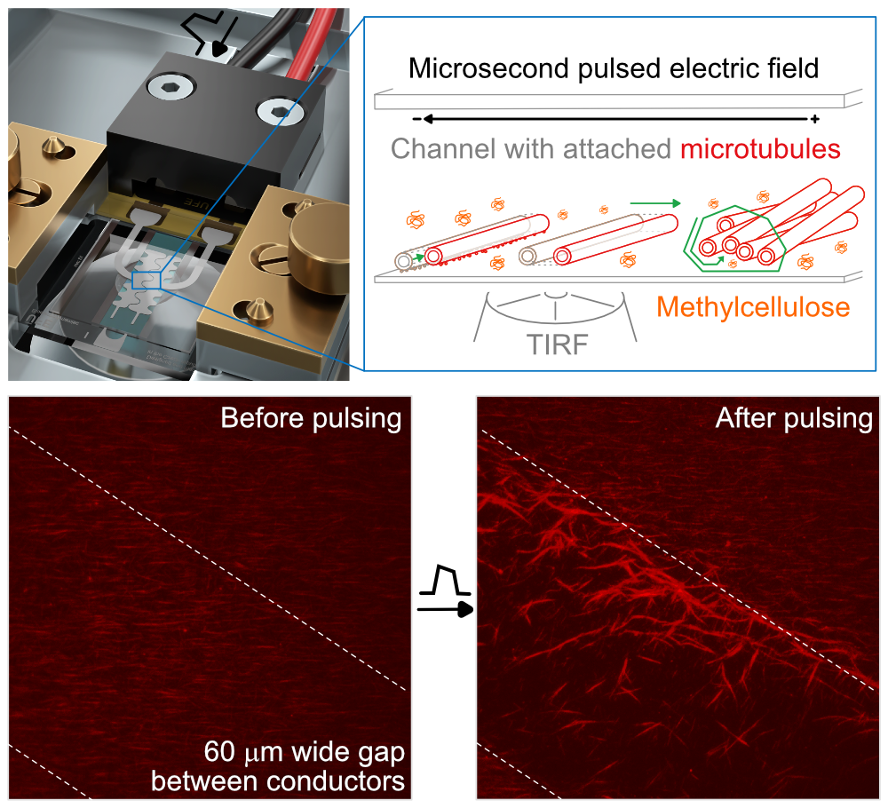
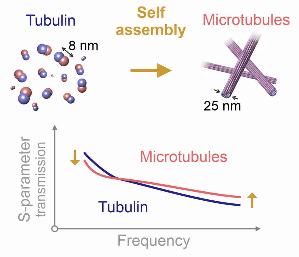
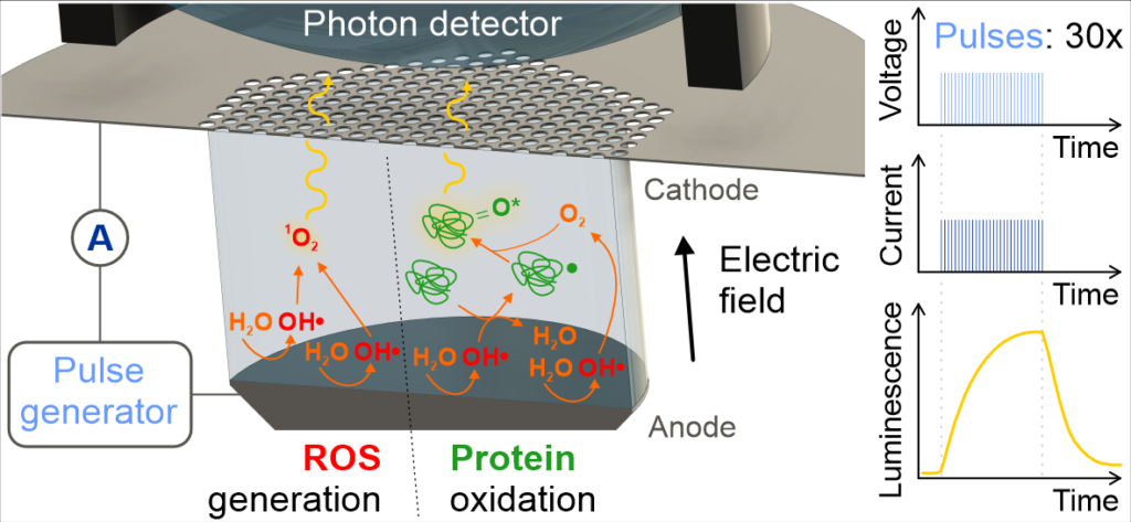
Teoretické modely a simulace ověřujeme skrze experimenty: v současné době se zaměřujeme na experimenty na nanostrukturách cytoskeletálních proteinů, konkrétně na mikrotubulech a s nimi souvisejících proteinech. Mikrotubuly jsou klíčové struktury nacházející se v každé buňce a umožňující dělení buněk a intracelulární transport. Naše současné projekty se zaměřují na měření elektromagnetických/dielektrických vlastností mikrotubulárních systémů a jejich řízení pomocí ultrakrátkých elektrických a elektromagnetických pulzů. Naše experimentální práce také zahrnuje měření a analýzu endogenního světla (luminiscence) buněk, v současné době pro monitorování oxidativních biologických vlivů elektromagnetického pole.

Publikace:

  1. Vahalová et al. “Biochemiluminescence Sensing of Protein Oxidation by Reactive Oxygen Species Generated by Pulsed Electric Field,” Sens Actuators B Chem, vol. 385, p. 133676, 2023.
  2. Poplová et al. “Biological Auto(chemi)luminescence Imaging of Oxidative Processes in Human Skin,” Analytical Chemistry, vol. 95, no. 40, pp. 14853–14860, 2023.
  3. Havelka et al. “Nanosecond Pulsed Electric Field Lab-on-Chip Integrated in Super-Resolution Microscope for Cytoskeleton Imaging.” Advanced Materials Technologies 5, no. 3 (2020): 1900669.
  4. Chafai et al. “Reversible and Irreversible Modulation of Tubulin Self‐Assembly by Intense Nanosecond Pulsed Electric Fields.” Advanced Materials 31, no. 39 (2019): 1903636.
  5. Krivosudský et al “Resolving Controversy of Unusually High Refractive Index of a Tubulin.” EPL (Europhysics Letters) 117, no. 3 (2017): 38003.
  6. Burgos et al. “Ultra-Weak Photon Emission as a Dynamic Tool for Monitoring Oxidative Stress Metabolism.” Scientific Reports 7, no. 1 (2017): 1229.
  7. Poplová et al. “Poisson Pre-Processing of Nonstationary Photonic Signals: Signals with Equality between Mean and Variance.” PLOS ONE 12, no. 12 (2017): e0188622.
  8. Saeidfirozeh et al. “Endogenous Chemiluminescence from Germinating Arabidopsis Thaliana Seeds.” Scientific Reports 8, no. 1 (2018): 16231.
  9. Sardarabadi et al. “Enhancement of the Biological Autoluminescence by Mito-Liposomal Gold Nanoparticle Nanocarriers.” Journal of Photochemistry and Photobiology B: Biology 204 (2020): 111812.
Skip to content