🚨From presentations to great conversations 💡
Michaela Poplová, Kateřina Červinková, and @mcer33 had a wonderful time at the #QuantumBiology Forum in Washington, with many inspiring and fruitful discussions along the way.


🛎️Conference season is on 🌍
Michaela Poplová, Kateřina Červinková and @mcer33 will represent our team next week in Washington at the #Quantum Biology Forum and #Optica Level Up.
Wishing them great conversations and exciting discoveries!💡👩🏫
🛎️Conference season is on 🌍
Michaela Poplová, Kateřina Červinková and @mcer33 will represent our team next week in Washington at the #Quantum Biology Forum and #Optica Level Up.
Wishing them great conversations and exciting discoveries!💡👩🏫
We stepped out of the office and into the brewery 🍺Learning, laughing, and bonding at @Plzen_Prazdroj -@Pilsner_Urquell brewery reminded us that strong teams are made from shared experiences—preferably well brewed ones. 🤩🥇 #TeamBuilding #PilsnerUrquell #BeerWithTheTeam 🍻
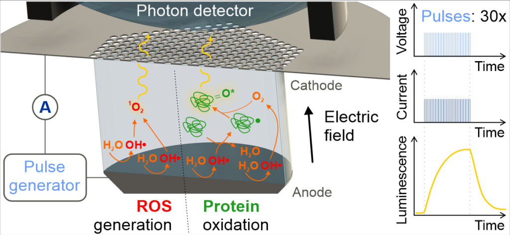
🔬⚡We introduce a calibrated, non-contact sub-THz probe station for microfluidic chips, delivering highly repeatable liquid and protein measurements across the F-band, with scalability toward 1 THz. Congratulations to @DanielHavelka5 and all the authors on this excellent work!👇
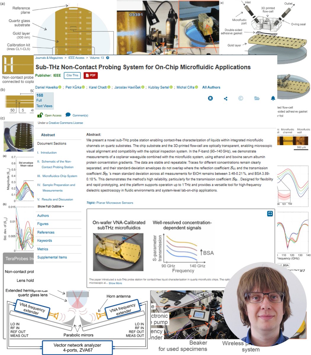

🔊🔬⚡We developed a sub-THz probe station for contact-free liquid sensing in transparent microfluidic chips. F-band tests show clear, stable signals—enabling reliable dielectric spectroscopy up to 1 THz.🔎📖👉https://ieeexplore.ieee.org/document/11153476